|
|
|
Home |
Program |
Speakers |
Meeting Registration |
Hotel Reservation |
Hotel Information |
Fellow Presentations
Musculoskeletal Ultrasound Evaluation of Hemarthrosis in a Patient with System Lupus Erythematosus (SLE) with Acquired Hemophilia A/Acquired Factor VIII InhibitorsCrisostomo R. Baliog, Jr., MD1, Anthony M. Reginato, MD, PhD2
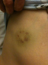
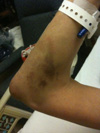
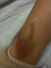
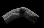
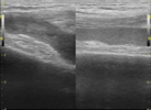
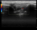
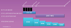
1Roger Williams Medical Center, Boston University School of Medicine, Providence, RI; 2Rhode Island Hospital, The Warren Alpert Medical School of Brown University, Providence, RI CaseA 47 F with SLE with membranous nephritis (Class V) presented to the ER with pleurtic chest pain and a three‐month history of ecchymosis on her extremities, anterior chest and face (Figure 1) without history of abuse or trauma. Chest pain spontaneously resolved and had no other clinical signs of lupus activity. Her medications at that time were Prednisone 15mg, Azathioprine 50 mg and Hydroxychloroquine. During her hospital course, she developed spontaneous swelling of the right elbow and knee. In order to determine the etiology of the oligoarticular swelling, a bedside musculoskeletal ultrasound of the right elbow and knee was performed in an attempt to evaluate the need for a diagnostic Arthrocentesis. Imaging StudiesMusculoskeletal ultrasound of the knee and elbow was performed showing complex areas of hypo and isoechogenecity on grey scale with minimal to no power‐Doppler signal. Synovial hypertrophy was noted in the affected knee compared with the unaffected knee. These complex images obtained in grey scale of the knee and elbow joints were then presumed to be from hemarthrosis. TreatmentTo stop progression of the hemarthrosis, activated Factor VII (NovoSeven) was administered. Prednisone was increased to 50 mg and oral cyclophosphamide 100mg was started. Because of the severity of the presentation and significantly high levels of Factor VIII inhibitors Rituximab 375mg/m2 was added weekly for four weeks. After 5 months of therapy, Cyclophosphamide was discontinued and Cyclosporine 100mg BID was started as maintenance therapy. Discussion
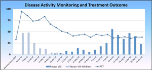
ConclusionMSKUS provided an inexpensive and real-time means to evaluate effusion and synovitis and served as a key imaging modality to diagnose hemarthrosis in an SLE patient with antibodies to Factor VIII and may potentially be used to monitor response to therapy. |
|
Administrative Office: Contact: Home |
Program |
Speakers | Meeting Registration |
Hotel Reservation | Hotel Information |
Fellow Presentations
|


